Diagnosis of Spinal Problems.


How to Diagnose Spinal Problems?
Diagnosis of spinal problems is necessary before starting any treatment. To arrive at a correct diagnosis, we need to perform appropriate Clinical examination, Have a complete Medical History.
Lots of Co-Existant Medical problems need to be properly managed, so that symptoms because of them do not overlap with Spinal Causes .
- Once Clinical diagnosis points towards a Spinal etiology, then specific investigations to confirm the diagnosis are carried out. Radiological Investigations like Xray of the affected area, MRI scan, and CT scan is done on outpatient basis , without getting admitted for the same.
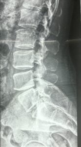
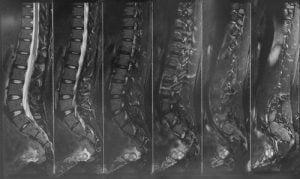
Figure showing Xray & MRI plate. A digital xray gives a proper image of the spine , while the MRI gives a detailed picture of the Spine with nerves and the spinal cord visualised clearly
- An MRI scan is the basic investigation required for diagnosis of a spinal problem as it shows the neural elements like the disc, the nerves and the spinal cord very well. A 1.5 T MRI scan is the minimum required scan nowadays, while some patients are claustrophobic and may find it difficult to go through the scan, for them the scan can be done under sedation, that is we give an injection so that patients can sleep through the procedure.
- Apart from an MRI other Radiological investigations like an Xray is also required. Its best to get a Digital Xray done, The quality and resolution is the best and this is important for the surgeon. The planning for a surgery also depends on a good quality X ray.
- A CT scan is an additional scan required in some cases when, there is suspicion of a tumour/infection or if the fracture pattern is not properly seen in X-rays & MRI. A CT scan gives better bony details than an Xray.
- Other Routine Investigations that are done are Dexa scan to check for bone quality and density, Blood Investigations to check for auto-immune pathologies and a Nerve study to check for nerve related pathologies.
Figure showing a dexa scan report which details the changes in bone density of the spine and hip bones.
- In cases where there is suspicion of spinal tumor or infection, A plain MRI scan may not be sufficient, we supplement it with a contrast MRI scan, where a contrast dye is injected , this helps in delineating between a tumour and other spinal pathologies.
- Apart from a contrast MRI scan, a PET SCAN or a Nuclear Bone scan is also done for cases where a tumour is suspected. These scans further help in determining wether the tumor has spread from some other organ, or it is originated from the spine only.
- When further confirmation is required, a CT guided or an open biopsy is advised to further confirm our diagnosis.
- Medical conditions like Diabetes, Thyroid disorders and Rheumatoid also need to be properly investigated. All these if present for a longer duration may lead to symptoms of back pain/neck pain, stiffness and spasms in arms or legs, since only MRI scans will by themselves not be enough, it is necessary to do appropriate blood investigations to rule out these causes.


